Viral culture
Below the techniques used for viral culturing are described
Background
Virus particles need mammalian cells to reproduce, which is why virus cultivation in the laboratory takes place in carefully selected host cells. The most commonly used cell type for, e.g., SARS-CoV-2, is the Vero cell, which is derived from the kidney of an African green monkey. This type is particularly suitable as it contains several of the surface properties necessary for a SARS-CoV-2 infection to occur. Additionally, this cell type shows clear signs of cell death if infected with SARS-CoV-2.

Detection of infectious virus
To assess whether a sample contains infectious virus, the sample is cultured in a culture flask with a large number of living cells. The sample is added to the culture flask, and the flask is left at 37°C for 3-7 days. If the cells show signs of death during this period, it indicates that a viral infection is occurring. The infection can then be confirmed by examining whether the amount of genetic material from the specific virus has increased over the course of the infection. Flask culture does not provide information about the quantity of virus in a sample, only whether the sample contains infectious virus or not.
Quantification of viral particles
Since viruses cannot be seen under a regular light microscope or replicate without a host cell, the amount of virus in a suspension is determined by measuring the number of dead host cells. One way to do this is by using a so-called plaque assay. Here, a patient sample or virus suspension is added to a dense layer of host cells, after which the cells are covered with a gel designed to ensure that the virus can only spread to neighboring cells and not uncontrolled to all cells in the flask. After 2-7 days, small areas of dead cells become visible by microscopy, and can be observedby the naked eye after staining.
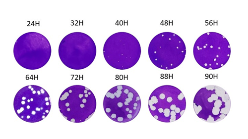
Picture showing plaque sizes in VERO E6 cells fixated 24-90 hours post infection with ancestral SARS-CoV-2 isolated in May 2020.
Assessment of viral neutralization capacity
To investigate how well a blood sample or a specific medication inhibits a particular virus type, a known amount of virus is mixed with different dilutions of the blood sample or medication. This allows for the calculation of the concentration at which 50% of the virus is neutralized, providing a measure of the effectiveness of a patient's blood or, for example, a laboratory-produced antibody. Neutralization of the virus means that the virus is either destroyed, it's binding to the host cell surface is prevented, or its replication mechanism intracellularly is inhibited, thereby blocking an infection.